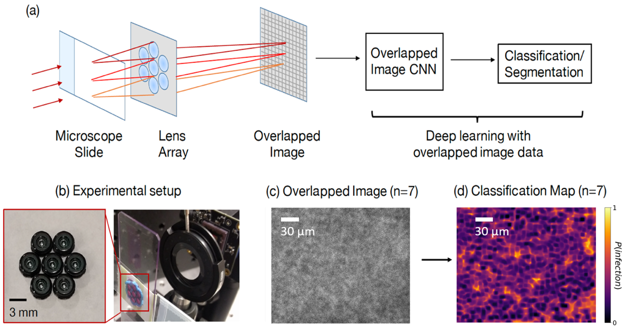
It is challenging and time-consuming for trained technicians to visually examine blood smears to reach a diagnostic conclusion. A good example is the identification of infection with the malaria parasite, which can take upwards of ten minutes of concentrated searching per patient. While digital microscope image capture and analysis software can help automate this process, the limited field-of-view of high-resolution objective lenses still makes this a challenging task. Here, we present an imaging system that simultaneously captures multiple images across a large effective field-of-view, overlaps these images onto a common detector, and then automatically classifies the overlapped image’s contents to increase malaria parasite detection throughput. We show that malaria parasite classification accuracy decreases approximately linearly as a function of image overlap number. For our experimentally captured data, we observe a classification accuracy decrease from approximately 0.9-0.95 for a single non-overlapped image, to approximately 0.7 for a 7X overlapped image. We demonstrate that it is possible to overlap seven unique images onto a common sensor within a compact, inexpensive microscope hardware design, utilizing off-the-shelf micro-objective lenses, while still offering relatively accurate classification of the presence or absence of the parasite within the acquired dataset. With additional development, this approach may offer a 7X potential speed-up for automated disease diagnosis from microscope image data over large fields-of-view.
Click here to visit the project page!